Unveiling the Secrets of Alzheimer’s Disease at the Nanoscale
Alzheimer’s disease (AD) is a debilitating neurodegenerative condition that affects millions of people worldwide. It is the leading cause of cognitive decline and death among seniors, accounting for about 70% of all neurodegenerative diseases. One of the hallmarks of AD is the accumulation of amyloid-β (Aβ) proteins, which form toxic aggregates known as amyloid plaques. To better understand the molecular mechanisms behind AD and develop effective treatments, researchers are continually exploring new techniques to study these proteins at the nanoscale.
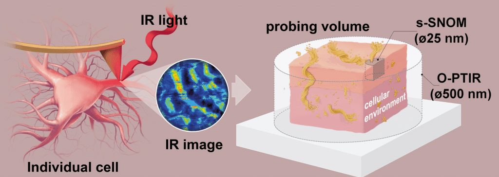
A breakthrough study led by an international team of researchers has demonstrated a new technique to detect Aβ-sheet structures in individual neurons without the need for immunolabeling. This innovative approach, called scattering-type scanning near-field optical microscopy (s-SNOM), allows scientists to analyze the structures at the nanoscale and gain a deeper understanding of the neurotoxicity caused by these aggregates.
Understanding Amyloid-β Proteins
Aβ proteins are associated with Alzheimer’s disease, and their accumulation in the brain forms amyloid plaques, which are toxic to neurons. Researchers believe that by eliminating these protein aggregates, they can slow the progression of the disease. However, current drugs targeting Aβ proteins have not been effective, indicating that the molecular mechanisms of Aβ neurotoxicity are not fully understood.
Traditional methods to study these proteins have limitations, such as insufficient spatial resolution or the need for prior knowledge of the targeted epitope structure for fluorophore labeling. To overcome these challenges, researchers have developed new cutting-edge technologies like s-SNOM, which offers high-resolution imaging at the nanoscale.
How s-SNOM Works
s-SNOM is a powerful imaging technique that combines atomic force microscopy (AFM) and infrared (IR) spectral imaging. It allows scientists to study amyloid structures at a spatial resolution of 20-30 nm, far better than what conventional microscopy techniques can achieve. This remarkable resolution allows researchers to identify and map molecular structures, enabling them to study protein aggregation in greater detail.
The Role of Ultra-Flat Gold
In this study, the researchers utilized ultra-flat gold from Platypus Technologies to create a conducive environment for high-resolution imaging of primary neurons. The gold-coated surface provided an ideal platform for the neurons to be cultured, enabling the researchers to investigate the Aβ-sheet structures directly in individual primary neurons using s-SNOM Nanoimaging and Nano-FTIR Measurements.
Key Findings and Future Implications
This groundbreaking research demonstrates the potential of s-SNOM as a valuable tool for neurobiologists to study amyloid structures at the nanoscale in neurons without the need for immunolabeling. The use of ultra-flat gold from Platypus Technologies played a crucial role in obtaining high-resolution images and valuable insights into the molecular mechanisms of Alzheimer’s disease.
As we continue to unravel the mysteries of Alzheimer’s disease, advanced technologies like s-SNOM, coupled with high-quality materials like ultra-flat gold, can help scientists better understand the disease’s underlying mechanisms. This knowledge could pave the way for more effective treatments and, ultimately, improve the lives of millions of people affected by Alzheimer’s disease.