How to Perform a Cell Migration Assay
Cell migration is a fundamental process in a variety of biological phenomena. This includes embryonic development, tissue regeneration, immune responses, and cancer metastasis. Understanding cell migration mechanisms is crucial for advancing therapeutic strategies in wound healing, immune therapies, and cancer treatment. Cell migration assays can help with the examination of the migratory responses of cells under various conditions.
This blog post delves into the methodology of conducting a cell migration assay. It focuses on the scratch assay, one of the most common and straightforward methods to assess cell migration in vitro.
Understanding Cell Migration
By comprehending how cell migrations work, we can learn more about their specific signals and the polarity of their signal molecules. That way we can hypothesize where they could move to. With the wrong signals come potential issues, including the spread of cancer cells.
The way a cell moves can be through different forces. Some simple cells use cilia or flagella. However, for more complicated cells, they could move through a change in a cell shape because of their cytoskeleton, and its focal adhesions, or any changes to cell junctions.
Cell migration assays are used to understand cell migration. This can involve cell culture media to conduct an examination and to encourage cell growth. 3D environments and microscopes can also be used to learn more about cell motility, including the cell’s leading edge and its actin filaments that move the cell forward.
Related: What are the Steps in Cell Migration?
Amoeboid Migration
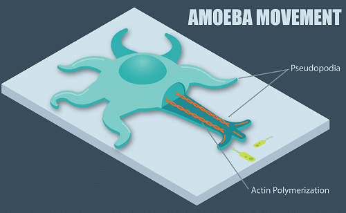
Another form of cell motility that you must understand is amoeboid migration. Instead of moving through an adhesive, their plasma membrane goes towards the back of the cell. The force of the plasma membrane can help it to be directed through the fluid that surrounds it.
You will mainly see this type of migration in immune cells. When tissue damage occurs, these cells need to be quickly directed to its source. Unfortunately, cancer cell metastasis also uses this type of movement.
Materials and Methods for Cell Migration Assay
Materials Required
- Cell culture: A cell line of interest that is well-characterized for in vitro migration studies.
- Culture medium: Appropriate for the specific cell line, supplemented with necessary growth factors, antibiotics, and serum, as required.
- 6-well or 12-well cell culture plates.
- Sterile pipette tips or scratch tools: For creating a uniform scratch.
- Inverted microscope: Equipped with a camera for capturing images.
- Image analysis software: To measure cell migration quantitatively.
Procedure
- Cell Seeding: Begin by seeding cells in a 6-well or 12-well plate, ensuring that they form a monolayer. The cell density should be optimized to reach confluence within 24-48 hours. This is critical for creating a uniform scratch.
- Establishing a Baseline: Once the cells reach 100% confluence, gently wash the cell layer with phosphate-buffered saline (PBS) to remove any debris and non-adherent cells. This step is crucial for obtaining clear and reproducible results.
- Creating the Scratch: Using a sterile pipette tip or a specialized scratch tool, create a straight scratch in the monolayer. Apply consistent pressure and speed to ensure the scratch width is uniform across different wells.
- Washing and Treatment Application: After scratching, gently wash the wells with PBS to remove detached cells. This reduces variability caused by floating cells. Following the wash, add fresh culture medium, with or without test compounds, to evaluate their effect on cell migration.
- Imaging: Immediately after creating the scratch (time 0), take photographs of the scratch using an inverted microscope. Ensure that the images are taken at standardized locations along the scratch to allow for accurate comparison over time.
- Incubation and Monitoring: Incubate the cells under standard conditions (37°C, 5% CO2). Monitor the scratch closure by taking images at predetermined time points, such as 0, 6, 12, 24, and 48 hours post-scratch. The time points may vary based on the cell type and the experimental design.
- Image Analysis: Analyze the images using image analysis software. Measure the area of the scratch at each time point. Then you can calculate the percentage of closure relative to the initial scratch area. This quantifies the cell migration rate.
Learn More: Alternatives to Scratch Assays
Results Interpretation
The rate of scratch closure reflects the migratory capacity of the cells. Enhanced migration will result in faster closure of the scratch. However inhibited migration will show a slower rate of closure. By performing multiple independent experiments, you can ensure statistical relevance and appropriate controls, especially when testing the effects of specific compounds on cell migration.
Limitations and Considerations
While the scratch assay is widely used because of its simplicity and low cost, it has limitations. The physical disruption of the monolayer can activate signaling pathways that do not reflect physiological cell migration. Additionally, this assay does not differentiate between cell migration and proliferation, which can both contribute to scratch closure. Advanced assays, such as transwell migration assays or three-dimensional migration assays, may be employed for more detailed studies.
Learn More: Comparison of the Oris Cell Migration Assay to the Scratch Assay
Are You Ready to Perform a Cell Migration Assay?
The scratch assay serves as a valuable tool in the study of cell migration. It provides insights into the basic mechanisms of cell movement and the effects of various substances on migratory behavior. With this knowledge, we can take further steps to control cell migration and learn the signs and results of specific signals.
Whether you are looking into dendritic cells or neural crest cells, by following standardized procedures and considering the assay’s limitations, researchers can obtain reliable data. This information can contribute to our understanding of cell migration in health and disease.
Mastering the intricacies of cell migration assays is paramount for researchers and professionals who are at the forefront of scientific discoveries in fields, such as oncology, immunology, and tissue engineering.
Platypus Technologies is dedicated to empowering your research endeavors with cutting-edge tools and innovative cell culture solutions. These are designed to enhance the accuracy and efficiency of cell migration studies.
Dive deeper into the world of cell migration assays and discover how our advanced technologies can streamline your research processes. Visit Platypus Tech’s Cell Migration Assay Guide to explore the possibilities and elevate your research to new heights.
Related: Unmatched Precision with Oris Cell Migration
References
- Guan JL, Liang CC, Park AY. In vitro scratch assay: a convenient and inexpensive method for the analysis of cell migration in vitro. Nature Protocols. 2007;2: 329-333. doi: 10.1038/nprot.2007.30.
- Amon J, Bartolini M, Cathcart J, Colarusso P, Jonkman J, Stevens K, Xu F. An introduction to the wound healing assay using live-cell microscopy. Cell Adhesion & Migration. 2014;8(5): 440-451. doi: 10.4161/cam.36224.
- Justus C, Leffler N, Ruiz-Echevarria M, Yang L. In vitro Cell Migration and Invasion Assays. Journal of Visualized Experiments. 2014;(88): 51046. doi: 10.3791/51046.