An Introduction to Wound Healing Assays
Wound healing assays are standard in vitro methods of probing collective cell migration in two dimensions. In wound healing assays, a cell-free area is formed in a confluent monolayer using physical exclusion or taking away the cells from the area via thermal, chemical, or mechanical damage. It is exposure to this cell-free area that leads to cells migrating into the gap.
Understanding Sheet Migration
Sheet migration is the type of collective cell migration that is probed by the wound healing assay. This migration is shown by endothelial and epithelial monolayers, traveling in two dimensions whilst still keeping their intercellular junctions.
Sheet migration can take place in a range of processes including cancer metastasis, tissue injury, and embryonic morphogenesis. There is a complex interplay within mechanical forces, biomedical cascades, and molecular interactions, and these are prompted by the exposure of the cellular monolayer to space when the cells make contact with the gap in the wound healing assay.
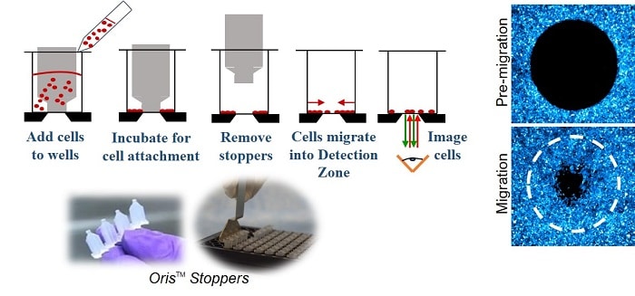
What Information Do Wound Healing Assays Provide
The information that is most commonly gained from wound healing assays is their gap closure rate. This is a measurement of the speed of the collective motion of the cells. In a standard experiment, gap closure rate is quantified under a range of conditions such as modulating the extracellular matrix composition, treating the cells with RNA interference, or changing a range of environmental variables including substrate stiffness.
Wound healing assays can also be adapted for medium and high throughput applications easily, such as drug discovery and small molecule screening.
Preparing Wound Healing Assays
Wound healing assays employ cells made from primary isolations from blood tissue or cell lines. Reproducible cell culture conditions are critical for stable phenotypes that are the basis of successful assays and conditions must be as reliable as possible.
Generally, wound healing assays are executed using a thin layer of cells that have been cultivated on a plastic or glass substrate. Most epithelial and endothelial cells have thin layers of cells that are obtained easily as they form confluent monolayers.
Wound healing assays should be introduced at the point at which the cells become confluent as the results gained may vary as the monolayer matures.
It is also important to consider the associated extracellular matrix when preparing wound healing assays. Some cell types can grow on plastic or glass substrates directly whereas some required a coating of collagen, gelatin, or fibronectin to adhere.
Learn More About Wound Healing Assays
Cell migration assay kits offer consistent detection zones to provide superior accuracy, reproducibility, and precision in experiments. Platypus Tech has a wealth of information on its website to help with your wound healing assay experiments. If you would like to find out more or discuss with an expert, visit the site today.