Description
ORIS™: A SIMPLE PLATFORM FOR PUBLICATION-READY DATA
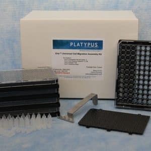
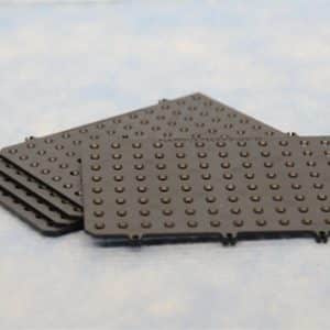
The Oris™ Cell Migration Assay platform uses a 96-well plate with “stopper” barriers that create a central cell-free Detection Zone for cell migration experiments. Removing the stoppers allows the cells to migrate into the Detection Zone at the center of each well.
BE CONFIDENT IN YOUR DATA
Oris™ Cell Migration Assay kits provide consistent Detection Zones to give you superior reproducibility, accuracy and precision in your experiments. The Oris™ Cell Migration Assay is the best option for publication-ready, high-quality data with fewer samples.
EXPERIMENT, FAST
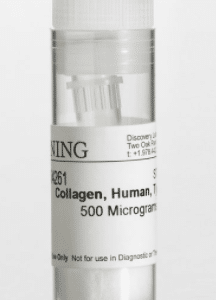
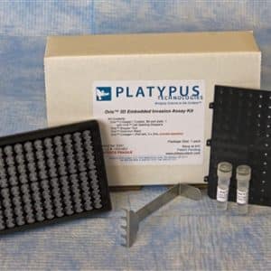
The Oris™ Cell Migration Assay kits can be used with any adherent cell line, and the wells of the assay are coated (Tissue Culture, Fibronectin or Collagen I) to support cell migration of the most common cell types. Additionally, the Oris™ Cell Migration Assay is compatible with plate readers and high-content analyzers, so you can rapidly quantify cell migration at any time during the duration of your experiment.
VERSATILE, TO SUIT YOUR NEEDS
Use the Oris™ Cell Migration Assay kits to identify cell migration inhibitors and cell migration promoters. For researchers in academia and pharmaceutical companies, the Oris™ Cell Migration Assay is a powerful tool to advance studies in drug discovery, wound healing, or cancer research.
Examples of Studies that used the Oris™ Cell Migration Assay:
- Y. Lee et al. “Downregulation of PRMT1 promotes the senescence and migration of non-MYCN amplified neuroblastoma SK-N-SH cells” Sci Rep 9, 177; 2019. LINK
- A. Kanda et al. “TGF-β-SNAIL axis induces Muller glial-mesenchymal transition in the pathogenesis of idiopathic epiretinal membrane” Sci Rep 9, 673; 2019. LINK
- M. Gnanamony et al. “Chronic radiation exposure of neuroblastoma cells reduces nMYC copy number” Oncology Lett. 14(3); 2017. LINK
- H. Marei et al. “Differential Rac1 signalling by guanine nucleotide exchange factors implicates FLII in regulating Rac1-driven cell migration” Nature Comm. 7; 10664; 2016. LINK
- S. Ravi et al. “Incorporation of fibronectin to enhance cytocompatibility in multilayer elastin-like protein scaffolds for tissue engineering” J. Biomed Mater. Res. A 101(7); 2013. LINK
- W. Gough et al. “A Quantitative, Facile, and High-Throughput Image-Based Cell Migration Method Is a Robust Alternative to Scratch Assay” J. Biomol. Screen. 16(2); pp.155-163; 2011. LINK
Related Products:
Single Plate Kits:
| Product Description | |
|---|---|
| Cell Migration Assay | Tissue Culture-Treated | 96-wells | BUY NOW |
| Cell Migration Assay | Collagen Coated | 96-wells | BUY NOW |
| Cell Migration Assay | Fibronectin Coated | 96-wells | BUY NOW |
Five Plate Kits:
| Product Description | |
|---|---|
| Cell Migration Assay | Tissue Culture-Treated | 5x96-wells | BUY NOW |
| Cell Migration Assay | Collagen Coated | 5x96-wells | BUY NOW |
| Cell Migration Assay | Fibronectin Coated | 5x96-wells | BUY NOW |