Description
Oris : A SIMPLE PLATFORM FOR PUBLICATION-READY DATA
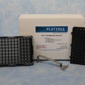
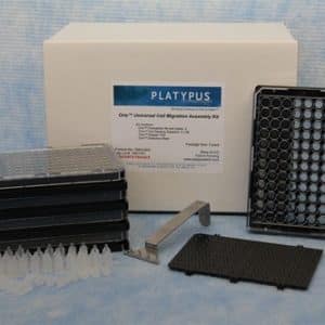
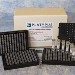
In the Oris 3D Cell Invasion Assay, stoppers are used to create a central collagen-containing Detection Zone for cell invasion experiments.
Cells are suspended in a thick layer of collagen surrounding a circular collagen Detection Zone. After incubation, invading cells move into the detection zone, where they can be unambiguously quantified using a microscope or a plate-reader.
Features & Benefits
- Monitor cell invasion entirely in 3D
- Oris platform enables you to distinguish different cell behavior in 2D cell migration and 3D cell invasion assay.
- Physiologically Relevant: No artificial membranes or inserts required.
- Versatile: Supports real-time monitoring or end-point analysis
- Easy Analysis: Invasion can be quantified using image analysis or plate readers; no elaborate cell tracking software needed.
Applications
Cell invasion assays are important tools for cancer research. Scientists studying cancer perform cell invasion assays to measure the cell movement through an extracellular matrix. By varying the conditions of the cell culture, scientists can identify conditions that prevent or accelerate cell invasion.
Examples of Studies that used Oris 3D Invasion Assay:
- J. Gu et al. “Targeting radiation-tolerant cells […]” Neuro-Oncology, 2022, LINK
- X. Zhang et al. “Actin cytoskeleton reorganization of hepatic stellate” Cell Adh Migr, 2019, LINK.
- L. Bertier et al. “Downregulation of invadopodium formation” Biomed. Pharm. 2018, LINK
- P. Masuzzo et al. “Analysis of high-throughput single-cell migration data” Sci. Reps. 2017, LINK